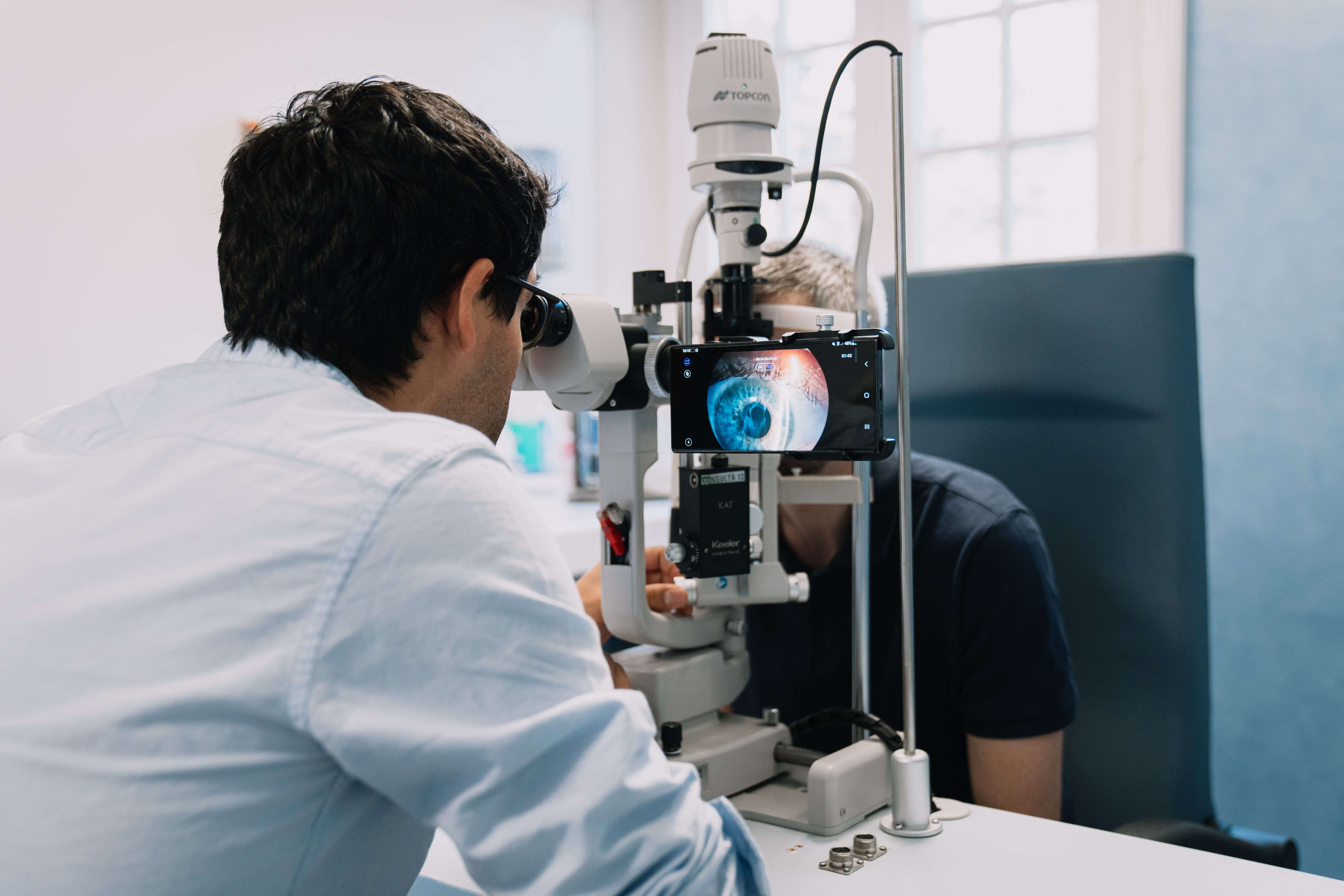
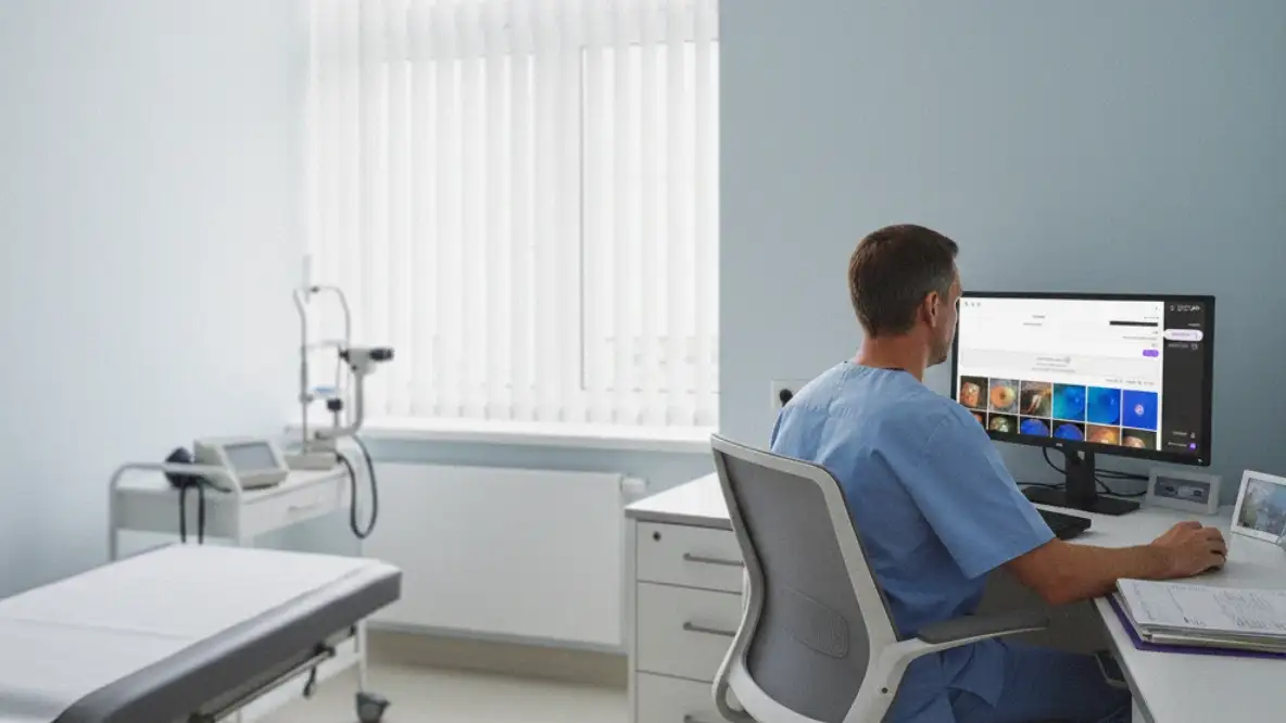
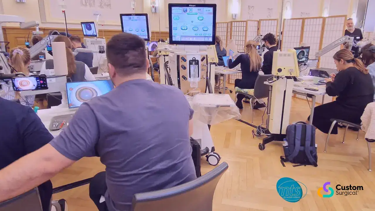
Smartphones have significantly impacted various aspects of our lives, including the field of medicine. Eye care professionals can now easily capture and share microscopic images during examinations, leading to improved diagnosis, treatment, and collaboration among specialists. Specialized optical systems, like MicroREC, make it simple to use smartphones for capturing microscopy images. By coupling these systems with ophthalmic instruments like slit lamps and surgical microscopes, eye care professionals can capture stable and clear images of the eye, maintaining stability during the process and ensuring an uninterrupted workflow.
Studies in ophthalmology have recently concentrated on testing various adapters and smartphones for slit lamp and microscopy imaging. A study conducted by German scientists compared 16 different smartphone models to determine their effectiveness in capturing images of the corneal endothelial cells (CECs). The smartphones were attached to the slit lamp eyepiece with an adjustable adapter, and healthy subjects were tested using the standard photo app of each phone to capture an image. 7 out of 16 smartphones could capture the CECs within 3 minutes. The study also recorded the subjective difficulty of taking a photograph and the factors contributing to successful image capture.

Source: Wiedenmann, C.J., Böhringer, D., Reinhard, T. et al. Korneale Endothelzellfotografie: Vergleich von Smartphones. Ophthalmologie 120, 382–389 (2023).
Exposure
The authors discuss that the correct exposure settings are essential for capturing the CECs. However, selecting the correct exposure setting can be challenging as CECs are only visible in medium light intensity. The automatic exposure of cameras often leads to overexposure of the dark areas, making it impossible to distinguish between CECs, or underexposure of the brightest areas, making the CECs appear too dark. Therefore, adjusting the exposure manually is necessary for most smartphones.
Our suggestion: Doctors who want to capture high-quality images of CECs can use the MicroREC App. It offers a solution to the challenges of achieving clear images by enabling users to manually adjust exposure settings and save them for future use. This feature eliminates the need to adjust exposure settings every time you use the app and prevents issues caused by automatic exposure, such as overexposure or underexposure.
Focus
It has been observed that the autofocus feature of certain cameras may not consistently focus on the CECs. This issue is further complicated because the slit lamp’s focus had to be guided through the smartphone’s display, as the adapter was attached to the eyepiece of the slit lamp. This resulted in unclear CECs caused by either a misaligned focal plane of the slit lamp or incorrect camera focus settings.
Our suggestion: The MicroREC is a convenient solution for capturing high-quality images without obstructing the slit lamp’s binoculars. By attaching to the slit lamp, it removes the need for camera autofocus, which can have difficulty focusing on the CECs consistently, or using a smartphone to guide the slit lamp’s focus, which can lead to misalignments and incorrect focus settings. This ensures reliable diagnostic results for ophthalmologists, resulting in better patient care.
Camera
Smartphones with both low and high camera resolutions (12 MP and 48 MP) could capture the CECs. However, a problem occurred because only one camera could be mounted in front of the eyepiece at a time due to smartphones switching between cameras when zoomed. As a result, it was impossible to quickly switch between cameras during exposure. Because of that issue, the authors conclude that smartphones with two cameras were more effective than those with three cameras in capturing the CECs.
Our suggestion: The MicroREC application provides doctors with a complete solution for utilizing smartphones with multiple cameras, as it manages and saves camera settings. The application controls which camera is being used for capturing images and videos, and saves the settings afterward. This eliminates the need to manually adjust smartphones on the eyepiece every time they’re used during a session, making the process quicker and more effortless.
In conclusion, the use of smartphones has revolutionized image capture and documentation in ophthalmology, resulting in improved diagnosis, treatment, and collaboration. Despite some challenges with exposure settings, autofocus inconsistencies, and camera selection, smartphones have proven to be invaluable for enhancing diagnosis, treatment, and collaboration among specialists. By using optical systems and apps like MicroREC and the MicroREC App that are specifically designed to improve the workflow of medical professionals, these issues are being addressed, leading to greater efficiency and accuracy in image capture. The utilization of smartphones ultimately leads to better patient care and improved visual health outcomes.
Source: Wiedenmann, C.J., Böhringer, D., Reinhard, T. et al. Korneale Endothelzellfotografie: Vergleich von Smartphones. Ophthalmologie 120, 382–389 (2023).


-1675d2368c6531b4186d0c38e40719e5.png)


