


-1675d2368c6531b4186d0c38e40719e5.png)


Do you document your success cases?
A patient came to me about a month ago, at the end of September, telling me that she had undergone cataract surgery in June, and since then, her vision had been poor. It seemed like she had a corneal decompensation and an ulcer.
When I examined her, at first, I thought it was indeed a case of corneal decompensation, with edema, which I found strange because the endothelium of her other eye’s cornea was in good condition.
But alright, it could have been a very traumatic cataract surgery, which I think was the case. That looked like this when on the first examination:

However, I found it odd that she had this ulcer without pain or any sensitivity, which made me think, “It can’t be!” The ulcer hadn’t healed since June
She had already tried numerous treatments, so I thought it might be herpes. I asked her, “Have you ever been treated for herpes?” and she replied, “No. The doctor even mentioned it could be bacteria, herpes, or a fungus.”
So, I decided to start her on herpes treatment, and the following week she returned with significant improvement. But the next week, her condition hadn’t improved further; it was similar to the previous week.

At the second consultation, I noticed that she had improved a bit, but something still suggested resistance to acyclovir. So, I switched to penciclovir, but when I saw her the following week, there was no progress at all.

Her daughter commented, “I think she followed the treatment properly in the first week, but not after that.” So, I changed the treatment, made a few more adjustments, and saw her today. Her cornea looks spectacular, like a different one!

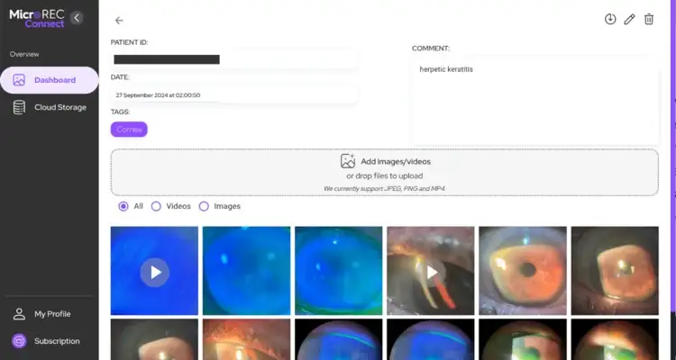
I managed to restore her cornea in a way I didn’t even think was possible, and all this progress is documented in Connect. You can see the entire improvement on Connect.

Which case are you proud to have documented? Share your story with us!
Author: Dr. Patricia Serapicos

Graduated in Medicine from the Faculdade de Ciências Médicas da Santa Casa de São Paulo in 2006 and completed her specialized residency in Ophthalmology at UNIFESP in 2010, focusing on Corneal Diseases and Refractive Surgery. With an additional specialization at the Ophthalmological Hospital of Sorocaba, she has extensive surgical experience in Corneal Transplants. She practices at CCO Jardins in Itaim Bibi, alongside her father and brother, both experienced ophthalmologists. Committed to excellence, Patricia volunteers at UNIFESP, sharing her expertise with medical trainees.